- +91-9646076888
- gauravmohan80@yahoo.co.in
- Amritsar, Punjab
Echocardiogram & Cardiac MRI
- Home
- Echocardiogram & Cardiac MRI
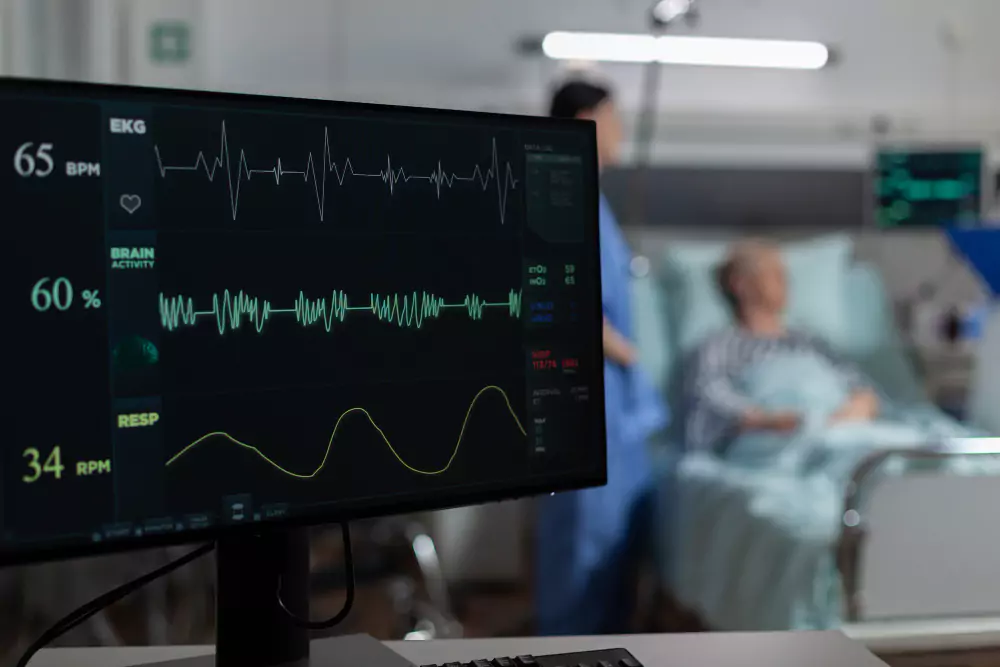
Echocardiogram (Echo)
An Echocardiogram is a non-invasive ultrasound test that provides detailed images of the heart’s structure and function in real-time.
✔ Indications: Recommended for patients with heart murmurs, valve disease, heart failure, chest pain, or suspected cardiomyopathy.
✔ Procedure:
A probe (transducer) is placed on the chest to emit sound waves.
The sound waves create detailed images of heart chambers, valves, and blood flow.
✔ Types of Echocardiograms:
Transthoracic Echocardiogram (TTE): Standard, non-invasive scan.
Transesophageal Echocardiogram (TEE): Provides clearer images using a probe inserted into the esophagus.
✔ Benefits:Assesses heart function, valve abnormalities, and structural defects.
Helps diagnose heart failure, clots, infections, and congenital heart disease.
Safe, painless, and radiation-free.
Cardiac MRI
A Cardiac MRI (Magnetic Resonance Imaging) is an advanced imaging technique that provides high-resolution, 3D images of the heart, offering superior detail compared to other scans.
✔ Indications: Used for detailed heart structure assessment, congenital defects, cardiomyopathy, myocarditis, and scarring from previous heart attacks.
✔ Procedure:
The patient lies in an MRI scanner.
A contrast dye may be injected to enhance blood flow and tissue imaging.
✔ Purpose:Provides precise evaluation of heart muscle, function, and blood supply.
Detects heart inflammation, fibrosis, or structural abnormalities.
✔ Benefits:Radiation-free, non-invasive, and highly accurate.
Helps in treatment planning for complex cardiac conditions.
With world-class expertise in non-invasive cardiac imaging, Dr. Gaurav Panchal ensures precise diagnosis and personalized heart care using the latest technology.
Book an appointment today for expert cardiac evaluation.